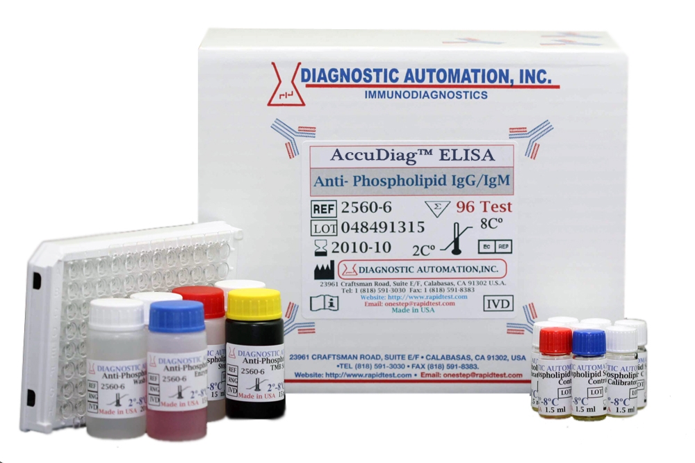
Name

Anti-Phospholipid ELISA test kit

Category Name Autoimmune Disease kits
Test 96
Method ELISA method: Enzyme Linked Immunosorbent Assay
Principle ELISA principle- Peroxidase conjugated
Detection Range 0-100U/mL IgG and IgM
Sample 100µl serum
Specificity Not Observed
Sensitivity 0.5U/mL
Total Time ~60 min
Shelf Life 12months

Item #:                    2560-6   Quantity:               

Anti-Phospholipid_2560-6_10-10-2009_.pdf
 

Anti-Phospholipid ELISA test kit


Description:


Anti-Phospholipid Screen (IgG/IgM) is an indirect solid phase enzyme immunometric assay (ELISA). It is designed for the quantitative measurement of IgG or IgM class autoantibodies directed against phospholipids. The microplate is coated with a mixture of highly purified negatively charged phospholipids: Cardiolipin, Phosphatidyl Serine, Phosphatidyl Inositol and Phosphatidic Acid. Anti-Phospholipid autoantibodies require Ã?2-Glycoprotein I as a co-factor for binding. The microplate is therefore saturated with human Ã?2-Glycoprotein I.


The microplates can be divided into 12 modules of 8 wells each or can be used completely for 96 determinations. Each well can be separated from the module ("break-away"). The binding of present autoantibodies, formation of the sandwich complexes and enzymatic colour reaction take place during three different reaction phases: (1) Calibrators, controls and prediluted patient samples are pipetted into the wells of the microplate. Any present antibodies bind to the inner surface of the wells. After 30 minutes incubation the microplate is washed with wash buffer for removing non reactive serum components. (2) An anti-human-IgG (or anti-human-IgM) horseradish peroxidase conjugate solution is pipetted into the wells of the microplate to recognize IgG class autoantibodies (or IgM class autoantibodies) bound to the immobilized antigens. After 15 minutes incubation any excess enzyme conjugate, which is not specifically bound is washed away with wash buffer. (3) A chromogenic substrate solution containing TMB (3,3`,5,5`-Tetramethylbenzidine) is dispensed into the wells. During 15 minutes of incubation the colour of the solutions changes into blue. Adding 1 M hydrochloric acid as stop solution stops colour development. The solutions colour change into yellow. The amount of colour is directly proportional to the concentration of IgG resp. IgM antibodies present in the original sample. To read the optical density a microplate reader with a 450 nm filter is required. Bichromatic measurement with a 600-690 nm reference is recommended.


The first study of anti-Phospholipid antibodies began in 1906, when Wasserman introduced a serological test for Syphilis. In 1942, the active component was found to be a phospholipid, which was designated Cardiolipin. In the 1950s it became clear that a number of people had positive tests for syphilis without any evidence of the disease. This phenomenon was referred to as the biological false positive serological test for syphilis. A high prevalence of autoimmune disorders, including systemic lupus erythematosus (SLE) and Sjögrens Syndrome occurred in this group of patients. The presence of circulating anticoagulants in patients with SLE was first documented in 1952 and was associated with increased risk of paradoxical Thrombosis in 1963. The term Lupus anticoagulant (LA), first used in 1972, is clearly a misnomer, because LA is more frequently encountered in patients without lupus and is associated with thrombosis rather than abnormal bleeding. During the last years it became clear that the optimal binding of anti-Phospholipid antibodies is depending on a cofactor termed Ã?2-Glycoprotein I (apolipoprotein H) (Ã?2GPI). Ã?2GPI is a 50-kDa b2-globulin occurring in plasma at a level of 200 μg/ml. It has been found that Ã?2-Glycoprotein I inhibits the intrinsic coagulation pathway and, therefore, it is involved in the regulation of blood coagulation. Ã?2GPI is associated in vivo with negatively charged substances, e.g. anionic phospholipids, heparin and lipoproteins. The phospholipid-binding region is located on its fifth domain. Under the acronym "aPL" (anti-Phospholipid antibodies) antibodies against negatively charged phospholipids, such as CL (Cardiolipin), LA (Lupus Anticoagulant), PS (PhosphatidylSerine), PI (Phosphatidyl Inositol) and PA (Phosphatidic Acid) are summarized. Of these, Cardiolipin is the phospholipid most commonly used as antigen to test for aPL by ELISA. Some Antisera, which bind cardiolipin-coated ELISA plates, can also bind to plates coated with other negatively charged phospholipids, such as Phosphatidyl Serine (PS), Phosphatidyl Inositol and Phosphatidic Acid (PA). Some investigators have suggested that the use of PS in place of cardiolipin in ELISA tests enables more specific diagnosis. These antigens are less commonly used and their additional use can improve the clinical sensitivity in patient samples with suspected APS (Anti-Phospholipid-Syndrome), but they can't replace the measurement of autoantibodies against Cardiolipin.  
Thyroid Elisa Kit
Categories Banner
Email Rapidtest
Order Elisa Kits
Order Elisa Kits