


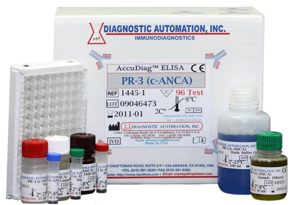
PR3 (c-ANCA) ELISA kit
| Name |
PR3 (c-ANCA) ELISA test |
|---|---|
| Full name |
Human PR3 (c-ANCA) ELISA Test Kit |
| Category Name | Autoimmune Disease kits |
| Test | 96 |
| Method | ELISA method: Enzyme Linked Immunosorbent Assay |
| Principle | ELISA - Indirect; Antigen Coated Plate |
| Detection Range | Semi-Quantitative: Positive, low positive, negative, cut off |
| Sample | 10ul |
| Specificity | 95.6% |
| Sensitivity | 98.2% |
| Total Time | ~75min |
| Shelf Life | 12 Months from the manufacturing date |
 |
 |
 |


PR3 (c-ANCA) ELISA kit description:
Diagnostic Automation Proteinase-3 (PR-3) ELISA is intended for the detection and semi-quantitative determination of antibodies to PR-3 in human sera. The assay is to be used to detect antibodies in a single serum specimen.
Material Provided with PR3 (c-ANCA) ELISA Kit:
1. Microplate: wells coated with Proteinase-3 antigen
2. Serum Diluent Type III
3. High Positive Control: Human serum or defibrinated plasma
4. Calibrator: Human serum or defibrinated plasma
5. Negative Control: Human serum or defibrinated plasma
6. Low Positive Control: Human serum or defibrinated plasma
7. HRP Conjugate: Goat anti- human IgG, IgA & IgM
8. Wash Buffer Type II:20X concentrate
9. Chromogen/Substrate Solution Type II: TMB
Materials Required, not Provided:
1. Precision pipettes
2. Distilled or deionized water
3. EIA kit Microplate Washer
4. EIA kit Microplate Reader with a 450 nm/dual 600-650nm
PR3 (c-ANCA) ELISA Kit Background Information:
Proteinase-3 (PR3)is a lysosomal enzyme that can be found in human neutrophils. There are two subsets of autoantibodies to human neutrophils: c- ANCA and p-ANCA. The subtype c-ANCA, shows cytoplasm staining by IFA and is diagnostic for Wegener granulomatosis. The major antigen responsible for the c-ANCA staining is PR3. PR3 is the most common antigen target of ANCA in patients with granulomatosis with polyangiitis where c-ANCA is found over 90% of the time. The second subtype p-ANCA, shows perinuclear staining by IFA. The major antigen responsible for the p-ANCA response has been shown to be myeloperoxidase. The ANCA staining patterns are obtained using ethanol-fixed human neutrophils. Anti- PR-3 autoantibodies have been associated with Wegener granulomatosis but less often with microscopic polyangiitis.
PR3 (c-ANCA) ELISA Test Principle:
PR-3 ELISA test is an Enzyme-Linked Immunosorbent Assay to detect IgG, IgA, and IgM antibodies to PR-3 antigens. Purified PR-3 antigens are attached microplet wells. Diluted test sera are added to each well. If the antibodies are present that recognize the antigen, antigen-antibody complexes are formed. After incubation, the wells are washed and an enzyme labeled anti-human IgG, A, M, is added to each well. If antibody is present the conjugate will bind to the antigen-antibody complexes. For additional details please refer to the instructions for use.
Notable features of Autoimmune Disease ELISA Kits:
User-friendly directions and explanation of test procedures
Simple and safe reagent preparation
Clear instructions on specimen collection
Comprehensive package of required materials
Explicit quality control and storage guidelines
Reliable and easy-to-read test results
Product inserts for all Autoimmune ELISA kits follow a similar method. See the PR3 (c-ANCA) ELISA Kit product insert for specific details on preparation, procedures, quality control, and test result interpretation.
Diagnostic Automation Inc. also provides other Autoimmune Disease ELISA Kits. For more information about ELISA Kits, Rapid Tests, IFA Kits, CLIA Test Kits, or Serology tests, please see our website home page, or contact our Customer Service Representatives at 818-591-3030 .


