


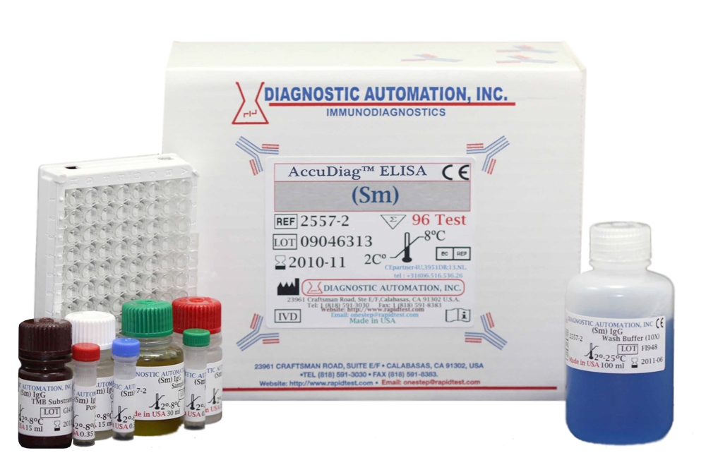
Sm ELISA kit
| Name |
Sm ELISA Test |
|---|---|
| Full name |
Human Sm ELISA Test Kit |
| Category Name | Autoimmune Disease kits |
| Test | 96 |
| Method | ELISA method: Enzyme Linked Immunosorbent Assay |
| Principle | ELISA principle - Peroxidase conjugated |
| Detection Range | Semi-Quantitative elisa assay- Positive, Negative and Cut-off |
| Sample | 10ul serum |
| Specificity | 99.3% |
| Sensitivity | 100% |
| Total Time | ~60 min |
| Shelf Life | 12 Months from the manufacturing date |
 |
 |
 |


Sm ELISA kit description:
Diagnostic Automation Sm ELISA test kit is a semi-quantitative immunoassay for the detection of IgG antibodies to Sm in human sera.
Material Provided with Sm IgG ELISA Kit:
1. Microplate: wells coated with inactivated antigen
2. Conjugate:HRP goat anti-human IgG
3. Positive Control: Human Serum
4. Calibrator: Human Serum
5. Negative Control: Human Serum
6. Sample Diluent
7. TMB
8. Stop Solution:1M H2SO4, 0.7M HCL
9. Wash Buffer Concentrate 10X
Materials Required, not Provided:
1. Precision pipettes
2. Distilled or deionized water
3. EIA kit Microplate Washer
4. EIA kit Microplate Reader with a 450 nm
Sm ELISA Kit Background Information:
The Jo-1 autoantibody is one of a family of characteristic autoantibodies seen in myositis patients. They are all specifically found in patients with myositis, and are all associated with a high incidence of accompanying interstitial lung disease. Antibodies directed against the Sm marker are highly specific for patients with SLE and are considered a diagnostic criterion for SLE. The presence of RNP antibodies in the serum of SLE patients is usually associated with a lower incidence of renal involvement and a more benign disease course. To the contrary patients with Sm antibodies experience a higher frequency of renal and central nervous system complications.
Sm ELISA Test Principle:
The Diagnostic Automation Sm ELISA test kit is designed to detect IgG class antibodies to Sm in human sera. Wells of plastic microwell strips are sensitized by passive absorption with immobilized antigens. The test procedure involves three incubation steps. For additional details please refer to the instructions for use.
Notable features of Autoimmune Disease ELISA Kits:
User-friendly directions and explanation of test procedures
Simple and safe reagent preparation
Clear instructions on specimen collection
Comprehensive package of required materials
Explicit quality control and storage guidelines
Reliable and easy-to-read test results
Product inserts for all Autoimmune ELISA kits follow a similar method. See the Sm ELISA Kit product insert for specific details on preparation, procedures, quality control, and test result interpretation.
Diagnostic Automation Inc. also provides other Autoimmune Disease ELISA Kits. For more information about ELISA Kits, Rapid Tests, IFA Kits, CLIA Test Kits, or Serology tests, please see our website home page, or contact our Customer Service Representatives at 818-591-3030 .
www.arabedroit.com