


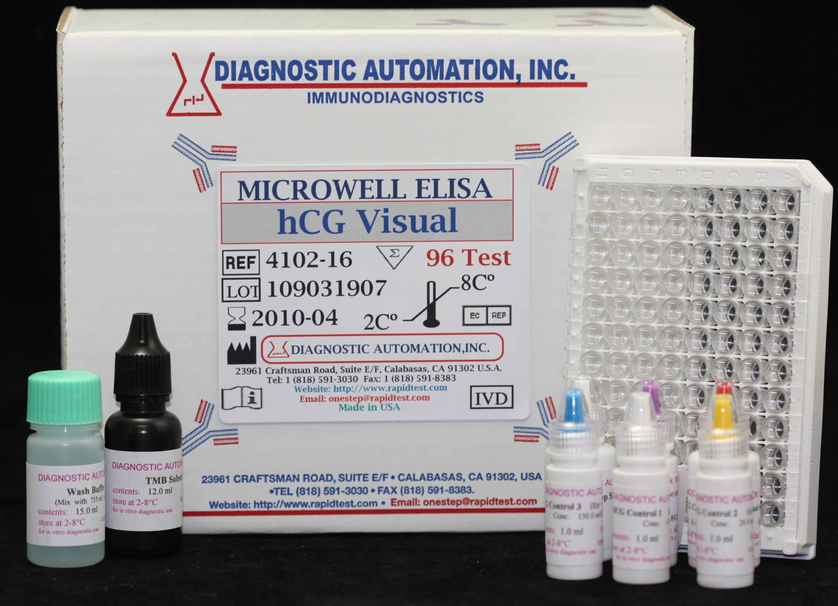
HCG Visual ELISA kit
| Name |
HCG Visual ELISA Test |
|---|---|
| Full name |
Human HCG Visual ELISA Test Kit |
| Category Name | Fertility ELISA kits |
| Test | 96 |
| Method | ELISA mehod: Enzyme Linked Immunosorbent Assay |
| Principle | ELISA principle- Peroxidase Conjugated Sandwich ELISA |
| Detection Range | 0-300mIU/mI |
| Sample | 10uI Serum |
| Specificity | 96% |
| Sensitivity | 1.0 ng/mL |
| Total Time | ~ 20 min |
| Shelf Life | 12 Months from manufacturing date |
 |
 |
 |


HCG Visual ELISA kit description:
The Diagnostic Automation hCG Visual EIA test kit is for the quantitative determination of Human chorionic gonadotropin (hCG) concentrations in human serum or urine.
Material Provided with hCG Visual ELISA Kit:
1. hCG Visual Elisa Test antibody coated Microtiter wells
2. Anti-hCG antibody HRPO Conjugate Reagent
3. hCG Control I 0.0 mIU/ml
4. hCG Control II 20.0 mIU/ml
5. hCG Visual EIA Kit Control III 150.0 mIU/ml
6. hCG Control IV 300.0 mIU/ml
7. TMB Substrate
8. hCG Visual Elisa Kit Stop Solution
9. Wash Buffer Concentrate50X
Materials Required, not Provided:
1. Precision pipettes
2. Distilled or deionized water
3. EIA kit Microplate Washer
4. EIA kit Microplate Reader with a 450 nm
hCG Visual ELISA Kit Background Information:
Human chorionic gonadotropin (hCG) is a glycoprotein hormone secreted by the developing placenta shortly after fertilization. In normal pregnancy, hCG can be detected in serum as early as 7 days following conception, doubling every 1.3 to 2 days. At the time of the first missed menstrual period, serum hCG concentration is about 100 mIU/ml, and peak levels of 100,000~200,000 mIU/ml are seen at the end of the first trimester. Thus making it an excellent marker for the early detection of pregnancy. Elevated serum hCG levels comparable to those observed in early pregnancy may also be associated with trophoblastic or nontrophoblastic neoplasms such as hydatidiform mole, choriocarinoma; therefore, the possibility of such diseases should be ruled out before a positive hCG result is considered diagnostic for pregnancy. The hCG Visual Test Kits is a rapid test to detect the presence of hCG in urine or serum specimens in a qualitative format.
hCG Visual ELISA Test Principle:
The hCG Visual Test Kit is based on a solid phase enzyme-linked immunosorbent assay. The assay system utilizes one anti-hCG antibody for microtiter wells immobilization and another mouse monoclonal anti-hCG antibody in the antibody-enzyme horseradish peroxidase conjugate solution. The test specimen serum or urine is added to the hCG antibody coated microtiter wells and incubated with the hCG antibody labeled with horseradish peroxidase conjugate. If hCG is present in the specimen, the hCG molecules will be sandwiched between the solid phase and enzyme-linked antibodies.. For additional details please refer to the instructions for use.
Diagnostic Automation Inc. also provides other Autoimmune Disease ELISA Kits. For more information about ELISA Kits, Rapid Tests, IFA Kits, CLIA Test Kits, or Serology tests, please see our website home page, or contact our Customer Service Representatives at 818-591-3030 .


 ELISA kit")
 ELISA kit")

