


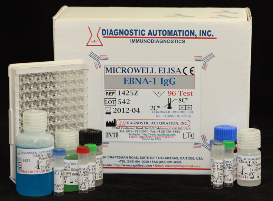
Epstein Barr Virus Nuclear Antigen (EBNA-1) IgG ELISA kit
| Name |
EBNA IgG ELISA Test |
|---|---|
| Full name |
Human Epstein Barr Virus Nuclear Antigen (EBNA-1) IgG ELISA Test Kit |
| Category Name | Infectious Disease ELISA kits |
| Test | 96 |
| Method | ELISA method: Enzyme Linked Immunosorbent Assay |
| Principle | ELISA principle- Indirect; Antigen Coated Plate |
| Detection Range | Qualitative elisa assay- Positive, Negative and Cut-off |
| Sample | 100ul |
| Specificity | 100% |
| Sensitivity | 97.8% |
| Total Time | 90min |
| Shelf Life | 12 Months from the manufacturing date |
 |
 |
 |


Epstein Barr Virus Nuclear Antigen (EBNA-1) IgG ELISA kit description:
Diagnostic Automation Epstein Barr Virus Nuclear Antigen-1 (EBNA-1) IgG Enzyme-linked Immunosorbent Assay (ELISA), is intended for the qualitative and semi-quantitative determination of IgG antibody in human serum to EBNA-1 recombinant antigen. EBNA-1 IgG assay may be used in conjunction with other Epstein-Barr tests.
Materials Provided with EBNA-1 IgG Elisa Kit:
1. Microwell strips: Recombinant EBNA-1 antigen coated wells
2. Serum Diluent Type I
3. Calibrator: human serum or defibrinated plasma
4. High Positive Control: human serum or defibrinated plasma
5. Low Positive Control: human serum or defibrinated plasma
6. Negative Control: human serum or defibrinated plasma
7. HRP Conjugate: Goat anti- human IgG
8. Chromogen/Substrate Solution Type I: TMB
9. Wash Buffer Type I: 20X concentrate
10. Stop Solution
Materials required but not provided:
1. Freshly distilled or deionized water
2. Dispensing system and/or pipette
3. EIA kit Microplate washer
4. EIA kit Microplate Reader with 450nm wavelength
EBNA-1 IgG Elisa Test Background Information:
EBV is classified as a member of the herpes-virus family based upon its characteristic morphology. EBV infection may demonstrate a wide spectrum of clinical symptoms. The majority of primary EBV infections is transmitted via saliva, occurs during childhood, and is subclinical. Antibody titers to specific EBV antigens correlate with different stages of IM. Both IgM and IgG antibodies to the viral capsid antigen (VCA) peak 3 to 4 weeks after primary EBV infection. IgM anti-VCA declines rapidly and is usually undetectable after 12 weeks. IgG anti-VCA titers decline slowly after peaking but last indefinitely. Antibodies to EBV nuclear antigen (EBNA) detected by anti-complement immunofluorescence develop from 1 month to 6 months after infection; and, like anti-VCA, persist indefinitely. Antibodies to EBNA indicate that the EBV infection was not recent. Antibodies to EA may appear transiently for up to three months or longer during the acute phase of IM in 85% of patients. Elevated levels of anti-EA and IgG anti-VCA may be detected in patients with chronic or recurrent illness suspected of being caused by EBV. However, a diagnosis of chronic EBV should not be based on the presence of antibodies to EA since elevated anti-EA titers may also be found in patients with other diseases as well as in healthy individuals with past EBV infections.
EBNA-1 IgG Elisa Test Principle:
Recombinant EBNA-1 antigen is coated on the surface of microwells. When antigens bound to the solid phase are brought into contact with a patient's serum, antigen specific antibody, if present, will bind to the antigen on the solid phase forming antigen-antibody complexes. Excess antibody is removed by washing. This is followed by the addition of goat antihuman IgG conjugated with horseradish peroxidase which then binds to the antibody-antigen complexes. The excess conjugate is removed by washing, followed by the addition of Chromogen/Substrate.
For more information about ELISA Kits, Rapid Tests, IFA Kits, CLIA Test Kits, or Serology tests, please see our website home page, or contact our Customer Service Representative at 818-591-3030.


IgG.jpg "Epstein Barr Virus Early Antigen (EA) IgG ELISA kit")


 ELISA test kit")
